Home |
Conditions |
Arm Pain
Thoracic Outlet Syndrome (TOS)
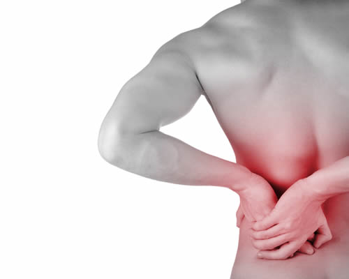
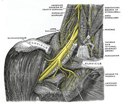
Thoracic Outlet Syndrome is caused by compression of the brachial plexus (main nerve bundle to the arm) and brachial vessels (artery and vein) in the root of the neck. It can produce a mixture of nerve compression symptoms in the arm and shoulder including pain, pins and needles, numbness, weakness, and circulation changes (sweating, blueness, blotchiness).
- Compression can occur in three separate places:-
Costoclavicular Syndrome occurs when the space between the collar bone (clavicle) and first rib is narrowed. This can be due to a congenital abnormality, or due to poor posture with rounded shoulders e.g. large breasts with a poorly fitting bra. - Cervical Rib Syndrome occurs when there is an extra rib or tight fibrous band connecting the 7th cervical vertebra with the sternum. The brachial plexus then has to pass over this extra structure causing nerve and vessel compression when the arm and shoulder are placed in certain positions. Symptoms most often occur down the inner arm to the little finger due to compression of the 8th cervical and 1st thoracic parts of the plexus.
Scalenus Anterior Syndrome (much rarer) occurs when the brachial plexus and vessels are trapped between the anterior and middle scalene muscles (similar to piriformis syndrome in the buttock).
Clinical Findings
- In slim patients the cervical rib can be felt just above the collar bone.
- Some patients have symptoms when the armed is pulled downwards, and others when the arm is elevated. Relief of the symptoms occurs when the arm is moved in the opposite direction.
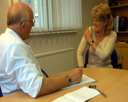
- Adson's Test is used to look for suspected compression. The patient looks to the affected side taking a deep breath. The examiner lifts the arm away to the side to 90 degrees, and notes whether the radial pulse disappears. However there are many false positives, as the radial pulse may disappear in normal people as the head of the humerus (upper arm bone) compresses the brachial vessels when the arm is taken beyond 90 degrees.
- Bruit - performing the tests above may cause compression of the subclavian artery, producing a bruit (murmur) audible with the stethoscope.
Investigations
- X-rays of the chest and neck may reveal a bony cervical rib but not a tight fibrous band.
- MRI scanning is the most useful investigation for revealing soft tissue anatomical abnormalities.
- Subclavian angiography (dye test) may confirm subclavian vessel compression.
- EMG (Electromyography) studies may confirm nerve dysfunction due to nerve compression.
Treatment
- Spinal Manipulation to the lower neck and upper thoracic spine, and attention to posture can increase the space between the clavicle and the 1st rib in costoclavicular syndrome. Breast reduction and a properly fitting bra is advised for some women with over-large breasts.
- Exercises to stretch the scalene muscles and related tight tissues can relief scalene muscle syndrome.
- Surgery is sometimes required to remove a cervical rib, part of the 1st rib, or rarely the lower part of the scalene muscles.
Shoulder
by John Chaffey last review April 2014